
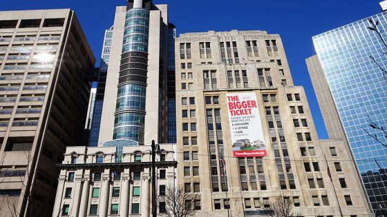
Princess Margaret Cancer Centre
Medical Imaging Leadership
Dr. Ur Metser
Site Director
Nancy Talbot
Interim Site Manager

Dr. Heidi Schmidt
Radiologist-in-Chief
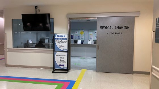
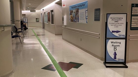
Location
3rd Floor, 610/620 University Avenue
Our Services
At Princess Margaret Cancer Centre we specialize in:
- Neurological imaging
- Cardiothoracic imaging
- Abdominal imaging
- Breast imaging
- Musculoskeletal imaging
- Molecular imaging
- Prostate imaging and biopsy
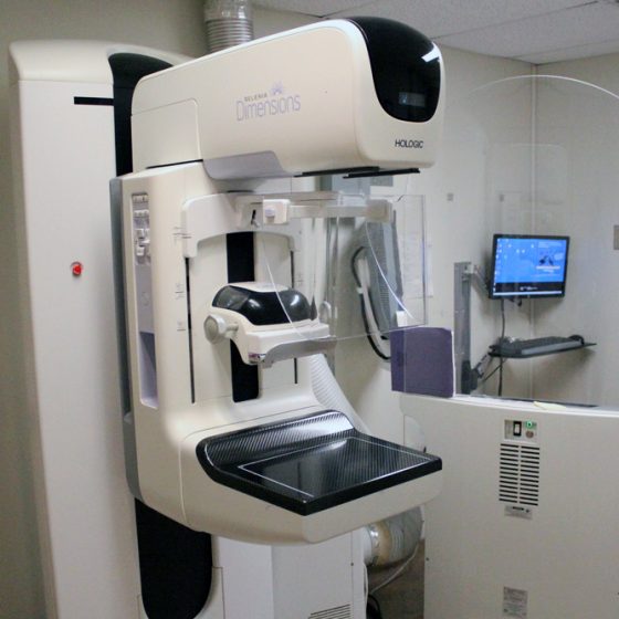
Breast Imaging – Breast Tomosynthesis (Type of Mammogram) and Mammogram
Hours of Operation:
Monday to Friday
7:30 a.m. – 5:00 p.m.
- Using X-rays and computer software, a breast tomosynthesis produces 3D images of the inside of your breasts.
- Low dose X-rays are used.
- Breast Tomosynthesis takes more images than a mammogram during the test.
Computed Tomography (CT)
Hours of Operation:
Monday to Friday
7:00 a.m. – 11:00 p.m.
- CT uses a pencil-thin X-ray beam to take detailed pictures inside the body.
- CT can be used to study all parts of the body.
- From CT images, colourful three-dimensional pictures of organs can be created.
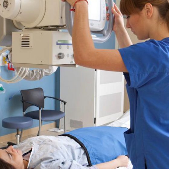
General Radiography (X-ray)
Hours of Operation:
Monday to Friday
7:00 a.m. – 6:00 p.m.
- X-rays are the oldest and most frequently used form of medical imaging.
- X-rays use small amounts of radiation to produce images of bones and internal organs.
- It’s non-invasive and helps physicians diagnose and treat medical conditions.
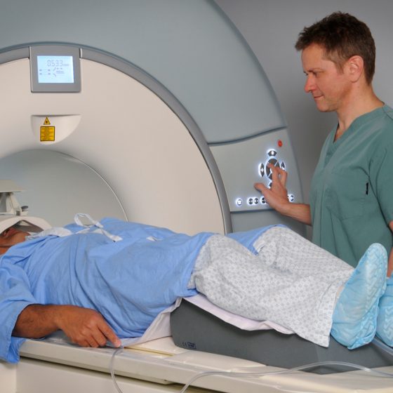
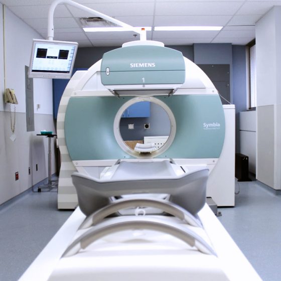
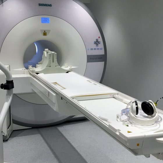
Magnetic Resonance Imaging (MRI)
Hours of Operation:
Saturday, Sunday, Monday
7:00 a.m. – 8:00 p.m.
Tuesday to Friday
7:00 a.m. – Midnight
Note: this may vary
- MRIs produce pictures of the body using powerful magnets and radio waves.
- MRI is a non-invasive procedure.
- Blood flow and organ motion can be measured with MRI
Positron Emission Tomography MRI (PETMR)
Hours of Operation:
Monday to Friday
8:00 a.m. – 6:00 p.m.
- PETMR combines technology from both molecular imaging and MRI.
- Images produced from injection of radiopharmaceutical are combined with MRI images.
- This new technology currently is used in research trials.
- PET scans your body using a small amount of radioactive sugar to help find certain diseases early.
- PET scans can also help show how well your treatment is working.
Prostate Center (4th Floor)
Hours of Operation:
Monday, Tuesday, Thursday, Friday,
8:00 a.m. – 12:00 p.m.
- Ultrasounds use sound waves to see the inside of the body in real-time.
- Biopsies take samples of tissue to assess tissue composition
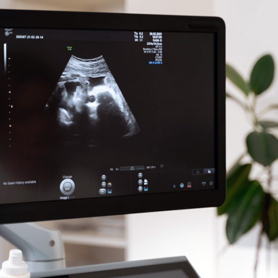
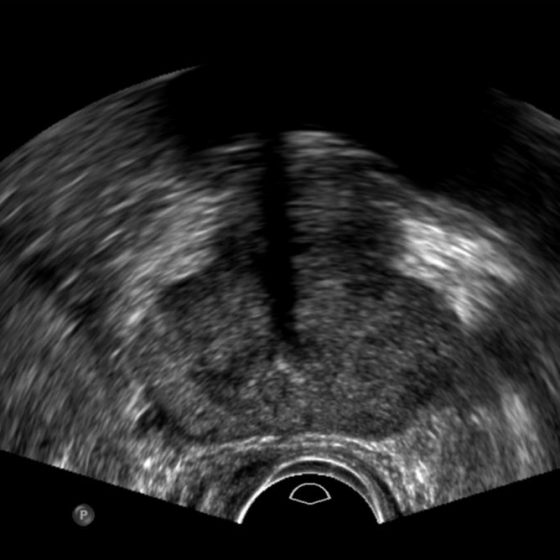
Ultrasound
Hours of Operation:
Monday to Friday
8:00 a.m. – 9:00 p.m.
- Ultrasound uses sound waves to see the inside of the body in real-time.
- It allows examination of organs, soft tissues and blood vessels.
- Common uses include examination of the uterus, liver, thyroid, heart—the list goes on!